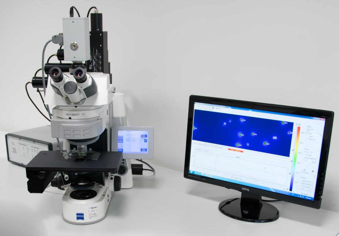


Fluorescence Kinetic Microscope FC 2000-Z
Fluorescence Kinetic Microscope FC 2000-Z (FKM) is designed to be the most versatile tool for lab-based research. Fluorescence Kinetic Microscope (FKM) extends the complete capacity of kinetic chlorophyll or multicolor fluorescence imaging to the realm of individual cells and subcellular structures. All conventional fluorescence parameters can be mapped with micrometer resolution so that individual chloroplasts or even grana-stroma thylakoid segments can be investigated. Modular set-up of the FKM is designed for kinetic fluorescence measurements with various user-selectable excitation and emission wavelengths as well as the combination of imaging measurements with spectrally resolved or ultra-fast (µs) spot measurements of fluorescence and absorbance kinetics.
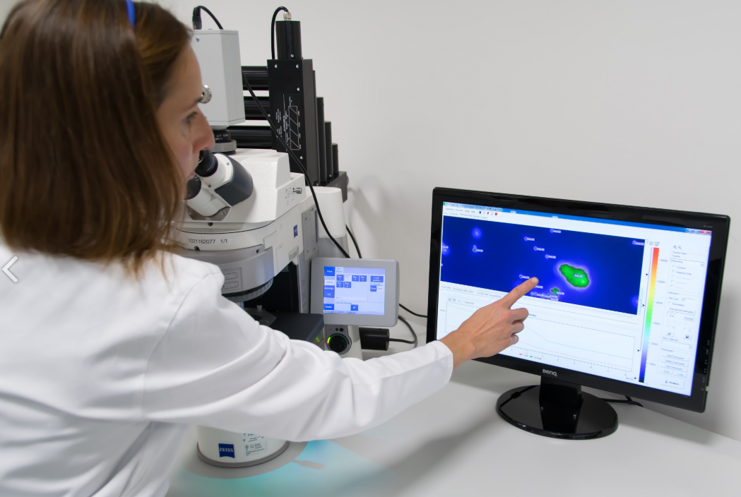
The Fluorescence Kinetic Microscope (FKM) allows imaging measurements of two-dimensional resolved multicolour fluorescence transients induced by complex irradiance protocols. The fluorescence emission is induced by an appropriate LED light source. In the standard configuration, the wavelength selection in Fluorescence Kinetic Microscope is the same as in other research-grade fluorescence microscopes by the use of a white excitation light source combined with a set of excitation filters, dichroic mirrors, and emitter filters. As an additional option, a light source with tunable spectra for both measuring and actinic / saturating light is available. It increases the range of measurable chromophores and improves signal/noise ratio. The system is designed to allow automatic switching between different excitation wavelengths even during same measurement. In contrast to conventional fluorescence microscopes, the ultrahigh sensitivity of the measuring camera combined with modulated light measurement makes it possible to perform imaging at extremely low light levels that do not disturb the metabolism of the cell.
The Fluorescence Kinetic Microscope can be combined with spectrally resolved measurements which are done via the Spectrometer SM 9000 synchronized to the measuring camera. The ultrafast kinetic measurements are performed by a microscope adapted version of the Double Modulation Fluorometer FL 3500, which again can be synchronized to the measuring camera. In this way, the spectrally resolved or ultrafast kinetic spot measurements can be done simultaneously on the same object and controlled by the same measuring protocol as the imaging kinetic record. All parts and functions can be controlled by the FluorCam software depending on individual configuration.
| Fluorescence Parameters |
Measured parameters: F0, FM, FV, F0', FM', FV', FT More than 50 calculated parameters: FV/FM, FV'/FM', PhiPSII, NPQ, qN, qP, Rfd, PAR-absorptivity coefficient, and many others |
|---|---|
| Light Sources | 455 nm, 470 nm, 505 nm, 570 nm, 605 nm, 618 nm, 630 nm, 735 nm, white and others |
| Super Pulse Intensity | 4,000 µmol. m-2. s-1 (in standard version) 6,000 µmol. m-2. s-1 (in light-upgraded version) |
| Actinic Light Intensity | Up to 2,000 µmol. m-2. s-1 (in standard version) Up to 3,000 µmol. m-2. s-1 (in light-upgraded version) |
| Filter Wheel | 7 positions |
| Light Regime | Static or harmonically modulated (sine waveform) |
| Custom-Defined Protocols | Variable timing, special language and scripts |
| CCD Detector Wavelength Range | 400 – 1000 nm |
| CCD Format | 720 x 560 pixels (TOMI-1 camera) 1360 x 1024 pixels (TOMI-2 camera) 1280 x 1024 pixels (TOMI-3 camera) |
| Pixel Size | 8.60 µm x 8.30 µm (TOMI-1) 6.45 µm x 6.45 µm (TOMI-2) 6.60 µm x 6.60 µm (TOMI-3) |
| A/D bit Resolution | 12 bit (TOMI-1) 16 bit (TOMI-2) 12 bit (TOMI-3) |
| Interface Connector | Gigabit Ethernet |
| Outer Dimension | 472 mm (W) x 479 mm (D) x 513 mm (H) |
| Weight | Appr. 40 kg |
| Power Input | Appr. 1100 W |
| Electrical | 90 - 240 V |
| Cameras | High-sensitivity TOMI-1 or high-resolution TOMI-2 or highspeed TOMI-3 |